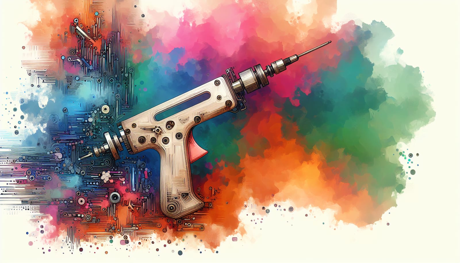
A remarkable advancement in medical technology has emerged with the development of a pioneering 3D printing “glue gun” that produces bone grafts right at the site of fractures. This innovative tool, created by dedicated researchers in South Korea, has the potential to transform surgical procedures by enabling the rapid creation of bone implants during surgery, eliminating the need for pre-made grafts.
This cutting-edge device is designed to fill in the gaps around the irregular edges of fractures, ensuring a perfect fit that promotes healing. The research team has diligently optimized the 3D-printed grafts, achieving a perfect balance of structural flexibility while also incorporating anti-inflammatory antibiotics that encourage natural bone regrowth at the grafting site.
Traditionally, bone implants have relied on metals, donor bones, or 3D-printed materials that require pre-surgical preparation. However, this new technology introduces a refreshing approach—a system that allows for real-time fabrication right in the operating room. As Professor Jung Seung Lee from Sungkyunkwan University explained, this innovation enables “highly accurate anatomical matching even in irregular or complex defects without the need for preoperative preparation.”
What’s truly fascinating about this “glue” is its composition. The filament used in the glue gun combines hydroxyapatite (HA), a natural component that aids healing, with a biocompatible thermoplastic known as polycaprolactone (PCL). The unique properties of PCL allow it to liquefy at relatively low temperatures, ensuring that tissue remains unharmed during application while perfectly conforming to the uneven surfaces of fractured bones.
The flexibility of the device is another highlight. Surgeons can easily adjust the printing direction and depth in real-time, allowing for quick and efficient graft creation during surgery. Professor Lee emphasized that this rapid process can be completed in just minutes, leading to reduced operative time and enhanced efficiency in surgical procedures.
Recognizing the importance of preventing infections, the researchers have thoughtfully integrated vancomycin and gentamicin into the filament. Their studies demonstrated that the scaffold effectively inhibited the growth of common bacteria that can lead to postoperative infections. The drugs are released gradually over several weeks, providing sustained protection at the surgical site.
In a successful proof of concept, the technology was tested on severe femoral bone fractures in rabbits, yielding excellent results. The team observed no signs of infection or tissue death after 12 weeks and noted enhanced bone regeneration compared to traditional grafting methods. The scaffold is designed not only to integrate seamlessly with surrounding bone tissue but also to degrade over time, making way for new bone formation.
The researchers are now focused on further enhancing the antibacterial properties of their scaffold and preparing for human trials. They are optimistic that this innovative approach will offer a practical and immediate solution for bone repair directly in the operating room, marking a significant leap forward in surgical care. This exciting development in medical technology exemplifies the power of innovation and collaboration in improving health outcomes.


